Johns Hopkins Researchers Fabricate Wireless Microgrippers for Grabbing Living Cells
- By Dian Schaffhauser
- 01/19/09
Johns Hopkins University researchers have invented dust-particle-sized devices that can be used to grab and remove living cells from hard-to-reach places without the need for electrical wires, tubes, or batteries. Instead, the devices are actuated by thermal or biochemical signals, paving the way for the use of tiny, mobile surgical tools activated by heat or chemicals. The mass-producible "microgrippers" measure about a tenth of a millimeter--one-254th of an inch--in diameter.
In lab tests, the tools have been used to perform a biopsy-like procedure on animal tissue placed at the end of a narrow tube. Experiments using the devices were reported recently by the National Academy of Sciences.
Although the devices will require further refinement before they can be used in humans, David Gracias, who supervised the project, said these thermobiochemically responsive, functional micro-tools represent a paradigm shift in engineering. "We've demonstrated tiny inexpensive tools that can be triggered en masse by nontoxic biochemicals," said Gracias, an assistant professor of chemical and biomolecular engineering in Johns Hopkins' Whiting School of Engineering. "This is an important first step toward creating a new set of biochemically responsive and perhaps even autonomous micro- and nanoscale surgical tools that could help doctors diagnose illnesses and administer treatment in a more efficient, less invasive way."
Presently, doctors who wish to collect cells or manipulate a bit of tissue inside a patient's body often use tethered microgrippers connected to thin wires or tubes. But the tethers can make it difficult navigate the tool. The untethered grippers devised by Gracias' team contain gold-plated nickel, allowing them to be steered by magnets outside the body. "With this method, we were able to remotely move the microgrippers a relatively long distance over tissue without getting stuck, he said. "Additionally, the microgrippers are triggered to close and extricate cells from tissue when exposed to certain biochemicals or biologically relevant temperatures."
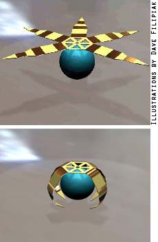
The microgripper, which resembles a crab, is fabricated using photolithography, the same process used to make computer chips. When the tiny devices are inserted in the body and moved magnetically, the gold-plated nickel in the palm and digits will allow doctors to see and guide the grippers with medical imaging units such as an MRI or CT.
In their lab experiments, the Johns Hopkins researchers used a microgripper, guided by a magnet, to grab and transport a dyed bead from among a group of colorless beads in a water solution. Team members also captured dozens of live animal cells from a cell mass at the end of a capillary tube. The cells were still alive 72 hours later, indicating the capture process did not injure them. Also, the microgrippers captured samples from relatively tough bovine bladder tissue.
Gracias' team is now working to overcome some remaining hurdles. As currently designed, each biologically compatible gripper can close on a target only once and can't be reactivated to reopen and release its contents. Gracias, who also is affiliated with the Institute for NanoBioTechnology at Johns Hopkins, hopes to collaborate with medical researchers who can help to move the microgrippers closer to use as practical biopsy and drug delivery tools in humans.
About the Author
Dian Schaffhauser is a former senior contributing editor for 1105 Media's education publications THE Journal, Campus Technology and Spaces4Learning.