Cornell Xray Imaging Lab Uses Kickstarter To Provide Low Cost Advanced 3D Imaging
Cornell University's 3D computed tomography (CT) imaging facility hopes to use crowd-funding platform Kickstarter to give non-invasive 3D, 4D and even 5D imaging to researchers everywhere.
Using as the theme of his campaign the phrase, "What would you do if you had Superman's X-ray vision?," Imaging Facility Director Mark Riccio said he hopes to raise $100,000 by Sept. 17.
With the funding, Riccio said he hopes to offer nonprofit organizations and individuals imaging at an hourly rate that is substantially lower than what standard imaging facilities charge.
| |
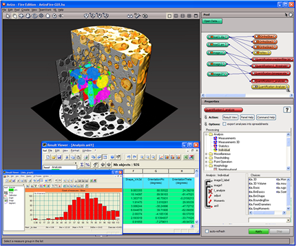
Mark Riccio wants to provide low-cost 3D CT imaging to more people and institutions, thereby creating a free online digital library of images. |
|
Riccio has divided users of Cornell's CT imaging services into two categories: "N-Peeps," for the nonprofits, and "F-Peeps," for those at for-profit organizations. It is the N-Peeps who would benefit from the lower rates Riccio hopes to supply with funding from the Kickstarter campaign.
Along with removing barriers to entry for N-Peeps, he also wants to create an online digital library of 3D data assets that will be gained through the use of the advanced imaging in hopes that it would add to the body of scientific knowledge shared openly on the Internet. The only exceptions to the free sharing would be proprietary information, which would not be imaged for lower costs.
With the $100,000 he hopes to raise in the next several weeks, Riccio wants to set up an independent imaging lab, acquire a state-of-the-art 3D X-ray imaging system and time on other CT instruments at other institutions to accommodate a broader range of samples.
The imaging system Riccio plans to acquire from Carl Zeiss X-ray Microscopy would be the most advanced submicron imaging system on the market today, allowing researchers to visualize and quantify inside their samples without destroying them. That would be particularly valuable in examining samples that could be altered or destroyed if they were cut open (like raw eggs) and rare or archival materials such as fossils or museum artifacts.
The Cornell X-ray CT lab already has imaged and has in its online digital library a variety of objects. Among them are a great white shark's head, fruits and flowers, coral, bearded dragons, the insides of electronics and advanced industrial materials.
About the Author
Michael Hart is a Los Angeles-based freelance writer and the former executive editor of THE Journal.